
P012 α4β7 Receptor occupancy by vedolizumab at trough time-point occurs at the membranal level and is not associated with endoscopic healing in inflammatory bowel disease patients
B. Ungar1,2, Z. Ben-Shatach1,2, O. Haj-Natour1,2, M. Yavzori1,2, E. Fudim1,2, O. Picard1,2, D. Coscas1,2, A. Lahat1,2, M. Lidar2,3, R. Eliakim1,2, U. Kopylov1,2, S. Ben-Horin1,2
1Sheba Medical Center Tel Hashomer, Gastroenterology Institute, Ramat gan, Israel, 2Sackler School of Medicine, Tel Aviv University, Tel Aviv, Israel, 3Sheba Medical Center Tel Hashomer, Rheumatology Institute, Ramat gan, Israel
Background
Previous studies have shown inconclusive associations between vedolizumab trough levels and therapy outcomes. A near-complete occupancy of blood and intestinal α4β7 was exerted by vedolizumab through a wide-range of drug levels, regardless of clinical outcomes. However, these observations were not conducted specifically at the time of trough drug levels, did not include α4β7 highly expressing eosinophils and did not establish whether occupancy is in fact due to fully internalised VDZ- α4β7 complex in the target T-cell.
Methods
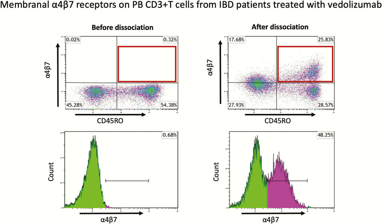
Peripheral blood (PB) and intestinal lamina propria (LP) were collected from IBD patients undergoing lower endoscopies on the day of vedolizumab infusion (trough time-point). Clinical and endoscopic scores were prospectively recorded. FACS analysis was performed using fluorescent conjugated-vedolizumab to investigate α4β7 occupancy on effector-memory T cells (CD3+CD45RO+ cells) and on eosinophils (CD49d+CD294+) in PB. Using a newly-developed dissociation assay, we also assessed the degree of membrane-bound VDZ-mediated blocked- α4β7.
Results
Forty-five IBD patients (16, 35% Crohn’s patients, 18, 40% in endoscopic remission) underwent clinical and endoscopic assessment just before their scheduled vedolizumab infusions. The median percentage of free α4β7 receptors at trough was 1.3% (IQR 0.5–3.7%) for PB T cells and 7.2% (IQR 3.6–23.4%) for LP T cells. No statistically significant difference was detected in α4β7 receptor saturation between patients with and without mucosal healing, both for PB (free α4β7 receptors: median 0.5%, 1.46%, IQR 0.3–1.4%, 0.9–3.5% respectively,
Conclusion
Even at trough, almost complete α4β7 receptor saturation is reached, both for PB and LP T cells, as well as for PB eosinophils, regardless of endoscopic activity status. This receptor occupancy is unrelated to receptor-internalisation and reflects a complete and continuous antigen-binding at the membranal level.