
P052 Nuclear magnetic resonance metabolomic profiling of IBD patients under anti-TNF treatment. Are the pathways network deregulated?
S. Notararigo1, M. Martin-Pastor2, J.E. Dominguez Munoz3, M. Barreiro-de Acosta3
1University Hospital Santiago De Compostela CHUS, Fundación IDIS, Santiago De Compostela, Spain, 2University Santiago de Compostela, Unidad de Resonancia Magnética, Cactus, Riaidt, Spain, 3University Hospital Santiago De Compostela CHUS, Department of Gastroenterology, IBD Unit, Santiago De Compostela, Spain
Background
The deregulation of immune system cell response implies loss of T-cell apoptosis, high rate of proinflammatory cytokines production and subsequent exacerbate activation of TNF-α pathway. The use of biologic antibody decrease inflammation rate and symptoms, but it remains unclear if it has a direct effect on the pathways activation/inactivation on peripheral blood mononuclear cells (PBMCs). The aim of this study is evaluate the role of nuclear magnetic resonance spectroscopy (NMR) applied to the metabolomic study of serum samples isolated from fresh blood from inflammatory bowel disease (IBD) patients under IFX treatment to understand the activated/inactivated pathways of PBMCs.
Methods
A case–control study was performed. Inclusion criteria were IBD patients under IFX treatment. Blood samples were obtained in Crohn’s disease (CD) and ulcerative colitis (UC) patients before IFX and in healthy controls (CTRL). CD patients were divided into subgroups according to the gut affected, in Ileocolic (IC), ileum and colon. NMR samples of the serum were collected and measured according to Standard Operation Procedures. Three types of NMR spectra were measured for each serum sample (1Hnoepresat, 1Hcpmgpresat and 1HDfilterpresat). The signal in each NMR spectrum was integrated in a series of equidistant little portion of the spectrum called buckets of a constant width of 0.04 ppm, covering the complete 1H NMR spectral window from −5 to 14 ppm. Buckets in regions depleted from signal at the two extremes of the spectrum were discarded as well as those in the proximity of the water peak at ca. 4.7 ppm which was affected by the presaturation. The vectors corresponding to a number of samples of two or more groups can be rapidly analysed using Multivariant Statistical Analysis methods.
Results
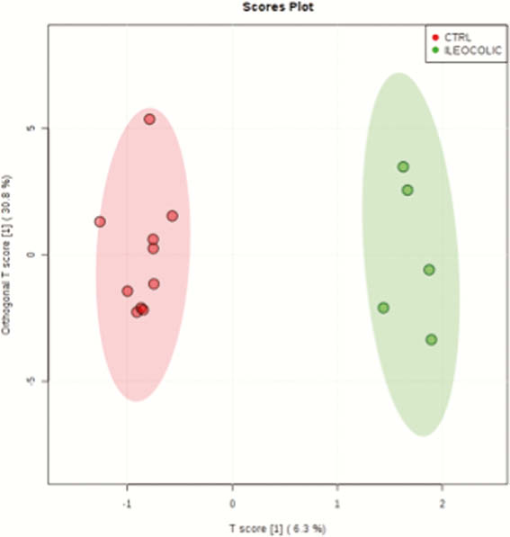
Twenty-two IBD patients (12 CD and nine UC) were included, 10 CTRL were also included. The metabolomic analyses of the NMR spectra of the serum of the different patients and control groups by the fingerprinting and targeting profiling strategies provided OPLS-DA statistical models (Figure 1) that permitted the successful classification of certain groups of samples which are summarised in Table 1.
| 1Hnoepresat | Fingerprinting | UC/CTRL |
| 1Hcpmgpresat | Fingerprinting | IC/CTRL, ILEUM/CTRL, COLON/CTRL |
| 1HDfilterpresat | Fingerprinting | IC/CTRL |
| 1Hnoepresat | Targeting profiling | UC/CTRL, IC/CTRL |
| 1Hcpmgpresat | Targeting profiling | IC/CTRL |
| 1HDfilterpresat | Targeting profiling | UC/CTRL, IC/CTRL |
Conclusion
The results of this pilot NMR metabolomic study of serum samples of IBD found a series of spectral fingerprints that are able to discriminate between groups of patients CTRL and CD, which underlines its potential use for the diagnosis of the disease.