P055 Effect of Mimosa caesalpiniifolia extract on DSS-induced colitis in Wistar rats

A. Garnevi Fávero1, T.S. Cordeiro1, K.N. Silva1, L. Cardili2, M.J.D. Silva3, A.P.R. Paiotti1

1Universidade Federal de São Paulo – Escola Paulista de Medicina UNIFESP/EPM, Discipline of Gastroenterology, Sao Paulo, Brazil, 2Universidade Federal de São Paulo – Escola Paulista de Medicina UNIFESP/EPM, Pathology, Sao Paulo, Brazil, 3Universidade Estadual Paulista, UNESP, São Vicente, Brazil, Biosciences Institute, Sao Paulo, Brazil

Background

Iinflammatory bowel disease (IBD) is a chronic disease characterised by dysregulation of the immune function response and imbalanced release of cytokines and unresolved inflammatory progress associated with the intestinal mucosa. In this way, new drugs are constantly tested as a novel treatment option. Mimosa caesalpiniifolia (MC) has been described as a potent analgesic, anti-inflammatory, anti-haemorrhagic, astringent, anthelmintic and diuretic. This study aimed to evaluate the effect of MC extract on dextran sulphate sodium (DSS)-induced colitis in Wistar rats.

Methods

Forty male Wistar rats were randomised into five groups: Sham; 5% DSS-induced colitis; control MC extract (125 mg/kg/day); 5% DSS-induced colitis treated with MC (125 mg/kg/day); 5% DSS-induced colitis treated with MC + 5-ASA (125 mg/kg/day for both). Colitis was induced by administration of 5% DSS (MP Biomedicals), diluted in drinking water and offered ad libitum for 5 days. MC plant extract was diluted in water and administered by oral gavage per 6 days. The leaf extracts were prepared in 70 gl ethanol and dried in a Buchi B19 Mini-spray dryer using 20% aerosol. The selection of the operating conditions used was followed by the Laboratory of Medicinal and Phytotherapeutic Plants of Unifal-MG. After the induction of colitis, body weight, general health status and faecal characteristics will be evaluated daily. All rats were euthanised on the ninth day of the experiment. The entire colon was dissected and fixed in 10% neutral-buffered formalin at room temperature and embedded in paraffin to provide sections for histological evaluation (Ethics Committee on the Use of Animals number 4362180119). The severity of colitis was evaluated in sections stained with Hematoxylin and Eosin. Parameters such as inflammation extent, regeneration and crypt damage were graded according to Dieleman et al. Statistical analysis was performed by one-way analysis of variance (ANOVA), followed by Tukey post hoc test using Graph Pad Prism (version 6.0).

Results

MC extract treatment reduced significantly the severity of DSS-induced colitis evidenced by a decreased in clinical symptoms (
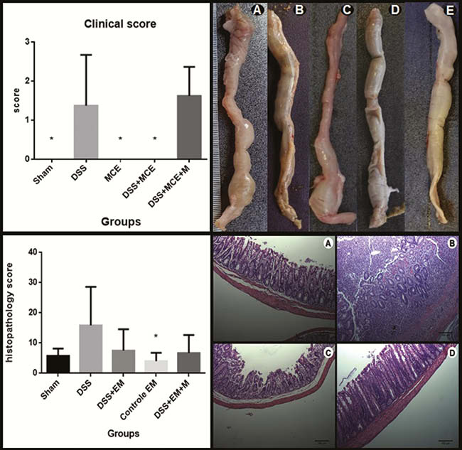
p < 0.0001). The rats treated with MC extract showed a decreased in diarrhoea episodes and rectal bleeding. We observed histopathological changes of colon tissue between the DSS-treatment and control groups, however, without statistical significance. The MC extract treatment reduced the damage to the colonic tissue evidenced by a decreased of inflammatory cells, ulcerations, and goblet cell preservation.

Conclusion

MC extract reduces the clinical signs and suggests a potential anti-inflammatory effect on DSS-induced colitis.