P056 Kefir (Lactobacillus kefiranofaciens) promotes autophagy and protects the intestinal barrier against DSS-induced colitis

K.N. Da Silva1, T.S. Cordeiro1, A.G. Fávero1, C.S. Bogsan2, R.D.C. Sinigaglia3, S.J. Miszputen1, O. Ambrogini–Júnior1, A.P.R. Paiotti1

1Universidade Federal de São Paulo – Escola Paulista de Medicina UNIFESP/EPM, Discipline of Gastroenterology, Sao Paulo, Brazil, 2Universidade de São Paulo – USP, Faculty of Pharmaceutical Sciences, São Paulo, Brazil, 3Universidade Federal de São Paulo – Escola Paulista de Medicina UNIFESP/EPM, Electron Microscopy Center, Sao Paulo, Brazil

Background

Inflammatory bowel disease occurs when luminal antigens from microbiota stimulate the immune system of an intestinal barrier through defects in the tight junctions lead to an exacerbated response in the genetically predisposed individual. It is well known that the consumption of probiotics improves intestinal health. Specifically, the Lactobacillus sp. strains play a key role in the epithelial intestinal barrier, promote mucin secretion, better performance of tight junctions and prevent apoptosis of epithelial cells. This study aimed to evaluate the role of Kefir (Lactobacillus kefiranofaciens) on inflammation of dextran sodium sulphate (DSS)-induced colitis.

Methods

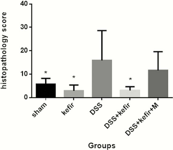
Forty male Wistar rats were randomised into five groups: Sham; 5% DSS-induced colitis; control Kefir; 5% DSS-induced colitis treated with Kefir; 5% DSS-induced colitis treated with Kefir + 5-ASA (125mg/kg/day) (Ethics Committee on the Use of Animals number 3627290119). The lactic culture of Kefir (Danisco Biolacta, Olstyn, Poland) was administered ad libitum per 6 days. The preparation of the matrix was carried out as follows: whole milk powder was reconstituted to 10% with distilled water and was incubated at 85°C for 15 min using a mechanical shaker. Subsequently, 20 mg freeze-dried Kefir culture was added to 100 ml of the treated milk and fermentation were carried out at 23°C for 16 h. When the desired pH of 4.6 was reached, the fermentation was stopped by cooling the flasks in ice bath and storing them under refrigeration at 4°C until utilisation. The rats were euthanised on day 9. Colonic samples were processed for transmission electron microscope (TEM) and conventional microscopy. Samples were analysed in a TEM EX II 1200 (JEOL, Japan) coupled to a digital camera system GATAN Orius (USA). Histopathological evaluation was performed by a pathologist under a light microscope. Parameters such as inflammation extent, regeneration and crypt damage were graded according to Dieleman et al.3 Statistical analysis was performed by one-way analysis of variance (ANOVA), followed by Tukey’s test using Graph Pad Prism (version 6.0).

Results

Kefir treatment reduces the severity of DSS-induced colitis evidenced by decreased abdominal discomfort and rectal bleeding. Histologically, there was a significant decreased of colonic damage and preservation of goblet cells (p < 0.0001) compared with DSS-colitis control group (Figure 1). The effect of Kefir was also verified by TEM analysis at the ultrastructural level, demonstrating the tight junctions preserved, decreased of reticulum edema and increased of autophagosomes (Figure 2).

Conclusion

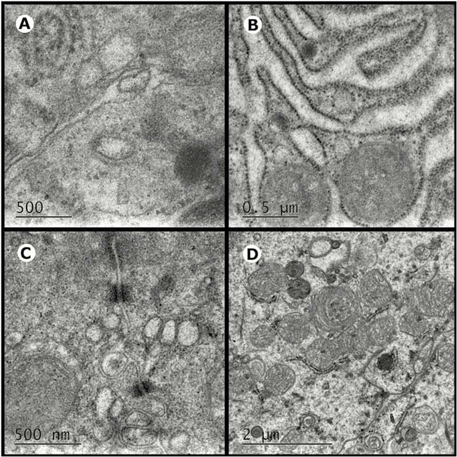
Kefir promotes autophagy and protects the intestinal barrier against damage caused by DSS.