
P269 Mucosal capillary pattern recognition based on real-time computer-aided image analysis adequately detects histological remission in ulcerative colitis
P. Bossuyt1,2, G. De Hertogh3, T. Eelbode4, S. Vermeire1, R. Bisschops1
1Departement of Gastroenterology and Hepatology, University Hospitals Leuven, Leuven, Belgium, 2Department of Gastroenterology, Imelda General Hospital Bonheiden,Bonheiden, Belgium, 3Departement of Pathology, University Hospitals Leuven, Leuven, Belgium, 4Medical Imaging Research Center, University Hospitals Leuven, Leuven, Belgium
Background
Objective treatment targets are required in a treat-to-target approach for ulcerative colitis (UC). An automated endoscopic system that correlates with histology can be an objective predictor for sustained remission in UC. The infiltration of neutrophils is associated with irregularities of the pericryptal capillaries. For this, we aimed to develop an objective automated endoscopic tool to assess histological remission based on the evaluation of the morphology of the pericryptal capillaries during endoscopy.
Methods
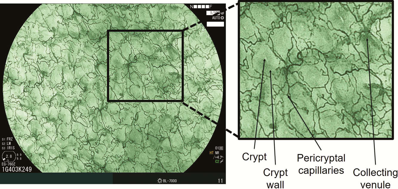
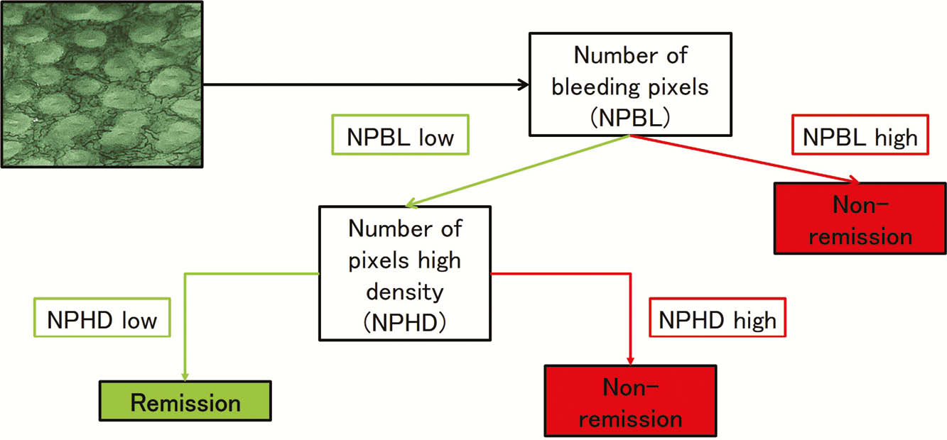
We used a prototype endoscopic system with short-wave-length monochromatic light from a LED system. This enables to evaluate in real-time the superficial (<200µm) mucosal architecture (crypts, pericryptal capillaries) and mucosal bleeding, Figure 1. An image analysis algorithm was applied to provide a score that quantifies the specific morphology of the mucosal capillaries. The algorithm included two steps. First, bleeding (mucosal/luminal) was assessed by pattern recognition. Samples with bleedings were automatically classified as non-remission. In case of non-bleeding, the degree of congestion of the capillaries was measured (maximal localised density estimation after morphological hessian based vessel recognition) to assess an ideal cut off value that identifies histological remission (Geboes score (GBS) <2B.1; no neutrophils in the lamina propria). Consecutive patients with UC were evaluated with the Mayo endoscopic subscore (MES), ulcerative colitis endoscopic index of severity (UCEIS) and the automated image analysis algorithm. To test the reliability of the algorithm and scores, the results were correlated with the GBS. Biopsies were taken in the matching area of the endoscopic evaluation.
Results
Fifty-eight patients with UC (53% male, median (IQR) age 41y (38–56), disease duration 7.1y (2.4–16.4)) with 113 evaluable segments (89% rectum or sigmoid) were included. The correlation between GBS and MES, UCEIS was good (
Conclusion
Mucosal capillary pattern recognition based on an automated image analysis with short-wave-length monochromatic light detects histological remission with high accuracy in UC. This technique provides an objective and quantitative tool to assess histological remission in UC, and excludes inter-reader variability.