
P443 Magnetic resonance imaging after ligation of the intersphincteric fistula tract for high perianal fistulas in Crohn’s disease: a retrospective cohort study
E. Van Praag1, K. van Rijn2, M. Monraats2, J. Stoker2, C. Buskens1
1Amsterdam UMC, Surgery, Vleuten, The Netherlands, 2Amsterdam UMC, Radiology and Nuclear Medicine, Amsterdam, The Netherlands
Background
Surgical closure of high perianal fistulas using the ligation of the intersphincteric fistula tract (LIFT) procedure is increasingly used in Crohn’s disease. Currently, data on MRI findings after the procedure is lacking, while this is the most important modality to assess deep fistula healing. Therefore, we aimed to evaluate pre- and postoperative fistula characteristics on MRI and the relation with clinical outcomes after LIFT procedure.
Methods
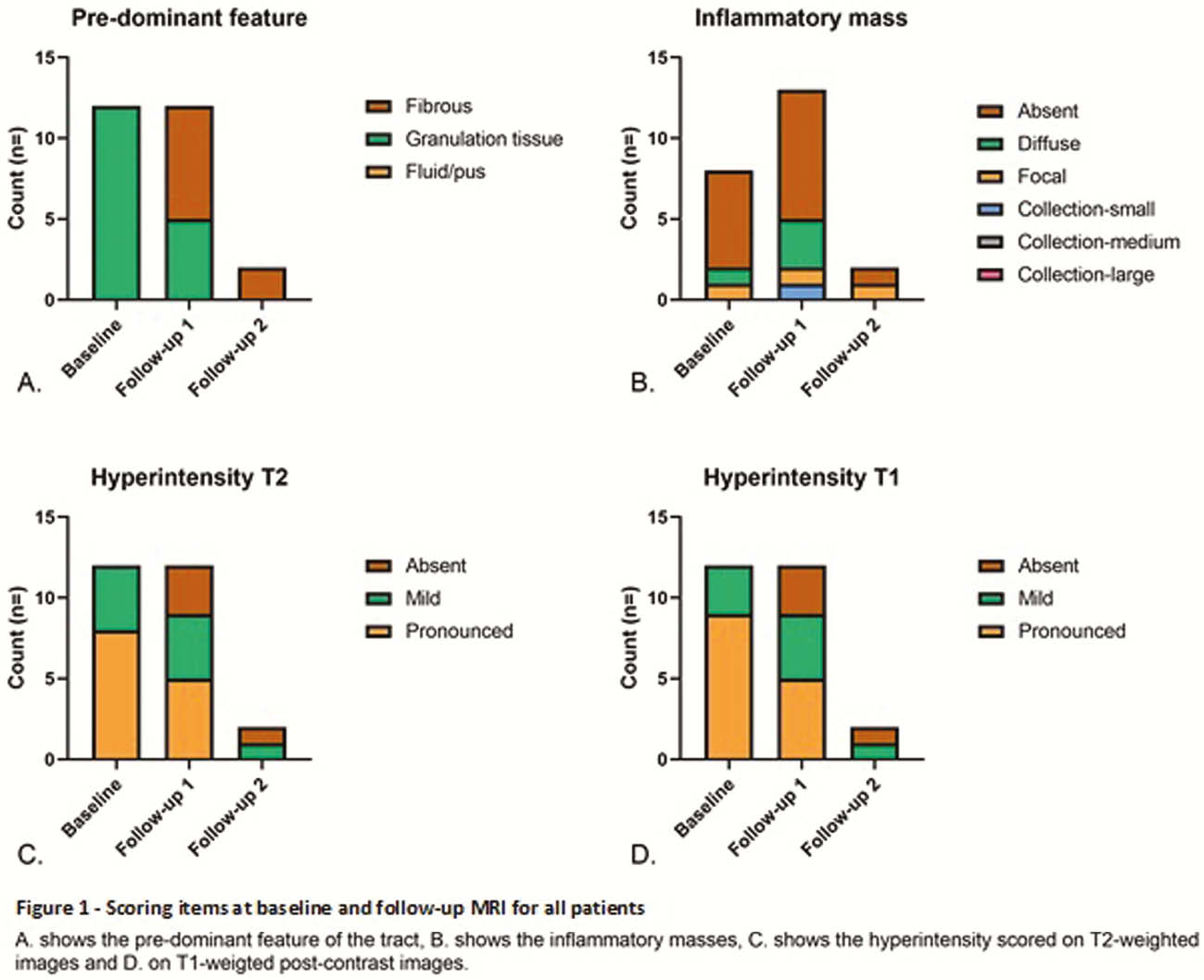
Consecutive Crohn’s patients treated with LIFT between 2007 and 2018 for high perianal fistulas who underwent a baseline and follow-up (FU) MRI were included in this retrospective study. MRI’s were scored by two radiologists according to a composed score based on the original and modified Van Assche scores with the addition of several items (Table 1). Findings at MRI and the relation to clinical healing were described.
| Number of fistula tracts | NoneSingle, unbranchedSingle, branchedMultiple |
| Hyperintensity T2 | AbsentMildPronounced |
| Changes in volume and/or hyperintensity on T2* | Substantial decreaseMinor decreaseComparableMinor increaseSubstantial increase |
| Inflammatory mass | AbsentDiffuseFocalCollection smallCollection mediumCollection large |
| Changes in inflammatory mass* | No massSame massNew mass (with location) |
| Hyperintensity on T1 | AbsentMildPronounced |
| Changes in volume and/or hyperintensity on post-contrast T1* | Substantial decreaseMinor decreaseComparableMinor increaseSubstantial increase |
| Dominant feature | FibrousGranulation tissueFluid/pus |
* These items were only scored on the follow-up MRI.
Results
A total of 12 patients were included (4 male, median age 34 years (IQR 28–39)). The FU MRI was performed a median 5.5 months (IQR 2.5–6.0) after the LIFT procedure. At this time eight patients (67%) reached clinical healing, which increased to ten patients (83%) during follow-up. None had a recurrence. Three patients (25%) needed a re-intervention after the FU MRI due to inflammatory masses and/or persisting fistula tracts. At baseline, all patients showed a tract predominantly filled with granulation tissue, which changed to predominantly fibrotic in seven patients (58%) (Figure 1). All clinically responding patients showed a decrease in tract volume and/or hyperintensity (i.e. activity) with an absence of hyperintensity on T1 and T2 in four (33%) patients.
Conclusion
Clearly decreased fistula activity can be observed on MRI after LIFT surgery in Crohn’s patients. A large proportion of patients develops a fibrotic tract relatively soon after the procedure and shows no clinical recurrences, suggesting a highly effective therapy and prognostic value of MRI.